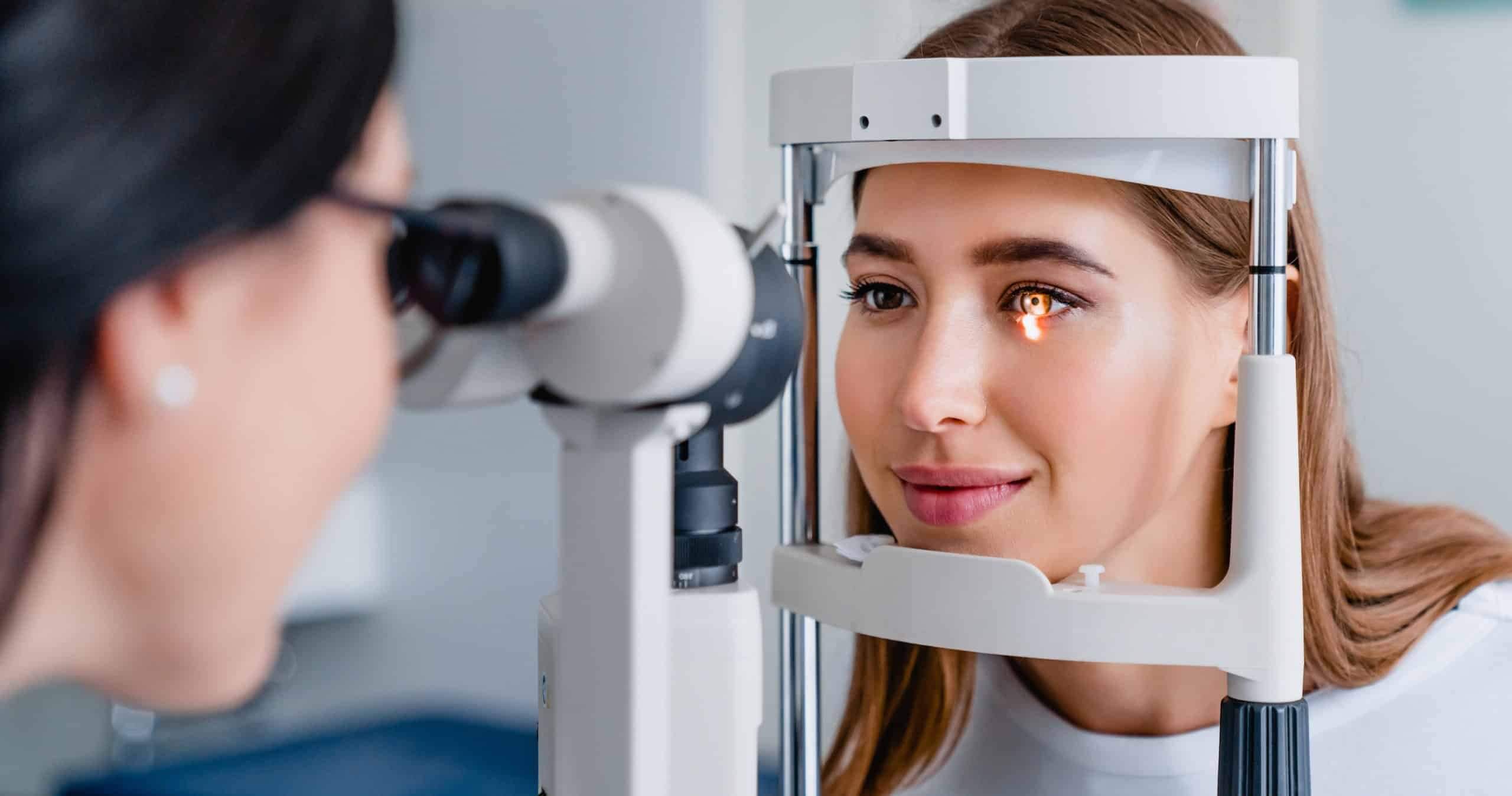
Ocular Coherence Tomography (OCT)
Optical Coherence Tomography or OCT is a high technology imaging test that allows the eye doctors at the Center for Sight to produce high resolution cross sectional views of the structures of your eye without ever touching it. The OCT test consists of multiple scans, and each scan takes about 45-60 seconds, the total test time is about 10 minutes. Since it is a non-contact, non-invasive testing method, it is not uncomfortable or difficult for patients. We perform Optical Coherence Tomography and its interpretation right in the comfort and convenience of our office at Center for Sight.
Optical Coherence Tomography (OCT) is particularly useful for studying Diabetic Retinopathy, Macular Edema, Macular Holes, Macular Pucker or Epi-Retinal Membranes, Macular Degeneration, Posterior Vitreous Detachment, Central Serous Retinopathy, Disorders of the Optic Nerve and even Glaucoma.


Fluorescein Angiography
A Fluorescein Angiogram (FA) or Intravenous Fluorescein Angiogram (IVF) is a diagnostic test that is used to study the retinal blood vessels and circulation of blood in the Retina. Fluorescein Angiography is a valuable test that provides information about many eye diseases including Diabetic Retinopathy, Macular Degeneration, Retinal Vascular Disease such as Retinal Artery Occlusion and Retinal Vein Occlusion as well as other types of Macular Disease.
Prior to beginning the study, your pupils will be dilated. The Fluorescein Angiography study is performed by injecting a sodium-based dye, called Sodium Fluorescein, into an arm vein.
During the injection, there can be a warm feeling or a hot flush. This only lasts seconds and then disappears. The dye appears in the retinal blood vessels within about 10-15 seconds. As the dye travels through the retinal blood vessels, an ophthalmic photographer or technician takes a series of photographs of the Retina with a special high-speed retinal camera. Capturing the photographs takes about 10 minutes.
If there are any abnormalities, the dye will usually reveal them by leaking, staining or by its inability to get through blocked blood vessels. Center for Sight’s Medical Retinal Specialist, Robert Kelly, M.D. will look for any abnormalities by identifying areas that exhibit hypo fluorescence (darkness) or hyper fluorescence (brightness). These are descriptive terms that refer to the relative brightness of fluorescence in comparison with a normal retinal angiography study.
Although statistically very rare, mild to severe adverse reactions to the intravenous dye have been reported. Dr. Kelly will review the potential risks and complications of Fluorescein Angiography with you and answer all of your questions prior to your study.
Visual Field Tests
A Visual Field Test is type of eye exam that that can identifies a loss of peripheral or “side vision”. A loss of peripheral vision may result from neurological problems such as stroke or tumors or eye problems such as glaucoma or retina disease. A Visual Field Test measures how far up, down, left and right you can see without moving your eyes AND how sensitive your vision is in various parts of the visual field.
The Visual Field Test is important for finding early signs of diseases like glaucoma that damage vision very gradually. Some people with glaucoma do not notice any problems with their vision, but the visual field test shows that peripheral vision is being lost. Further, the Visual Field Test examines the parts of the nervous system and brain that allow us to have vision and sight. The visual part of the nervous system includes the retina (the “film” in the camera-like eye), the optic nerve (the “wire” that carries images from the retina to the brain), and the brain itself. Problems with any part of this system can change the visual field. There are well-known patterns in the test results that help us recognize certain types of injury or disease. By repeating more visual field tests at regular intervals, we can also tell whether someone is getting better or worse.

At Center for Sight our technicians perform visual field tests with an advanced computerized digital instrument that allows a great deal of precision and allows us to store your testing for future comparison.